


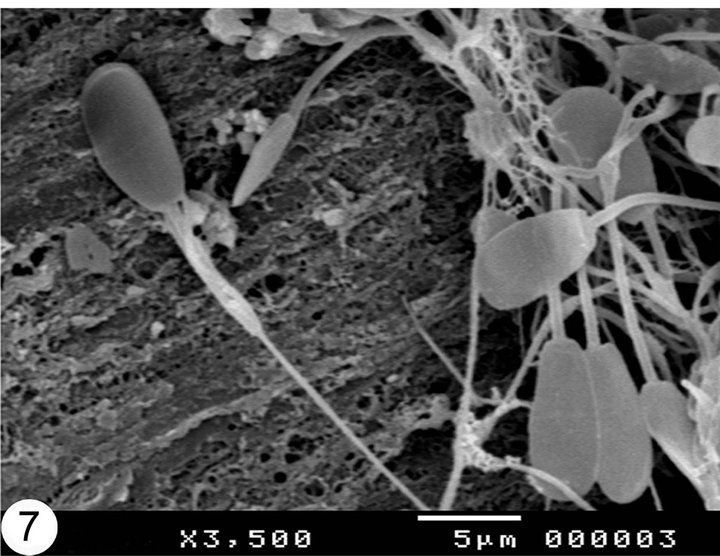
Micrographs of sperm


Adapted methods for scanning electron microscopy (SEM) in assessment of human sperm morphology


Want to find a partner you like Penetration? Find


Figure 3: Electron micrographs of sperm necks and tails. a The cross section of sperm tail from control group. b The ODF was lost in the sperm tail of exposure.


Figure 3. Electron micrographs of human sperm cells autometallographically developed for zinc ions. (A) Zinc grains are associated to the acrosome (ac), the .

There was a problem providing the content you requested


Description:Plate A is a light photomicrograph of sperm smear and green sperm heads in plate B show damage to sperm DNA under fluoroscent microscopy. Fluorescence micrograph showing the chromosomes from developing sperm cells. Nuclear transfer in Mouse Oocytes:
Views: 6001
Date: 2018-11-07
Favorited: 72


User Comments 1
Post a comment
Comment: